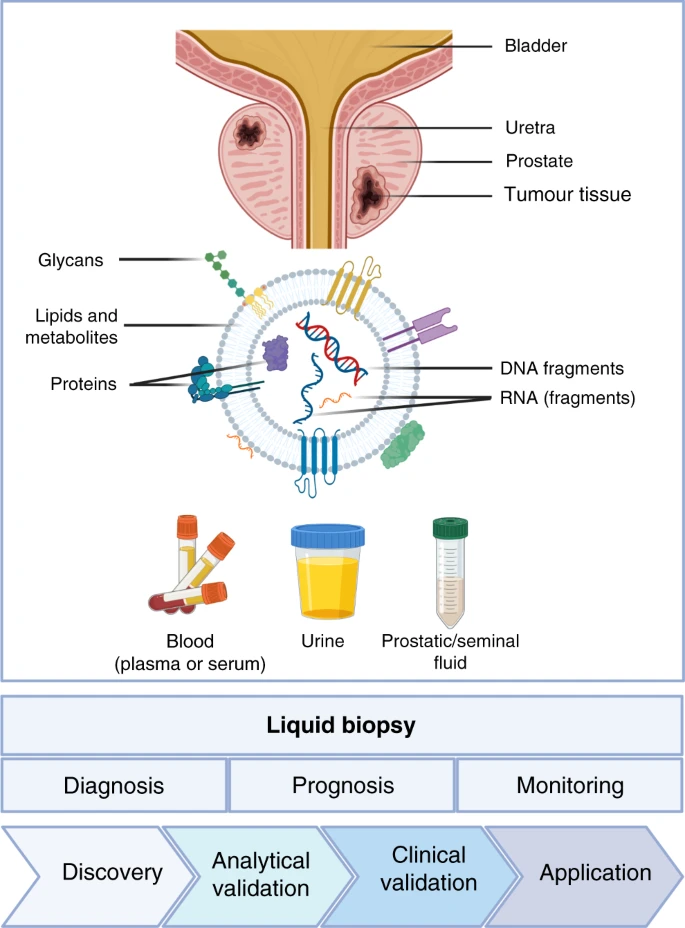
Liquid biopsy is a non-invasive method for examining biomarkers released into the bloodstream by tumors, such as cell-free nucleic acids (cfNAs), circulating tumor cells (CTCs), and extracellular vesicles (EVs). A fluid biopsy can possibly further develop malignant growth conclusion, forecast, and treatment by giving ongoing data about growth heterogeneity, advancement, and reaction to therapy [1].
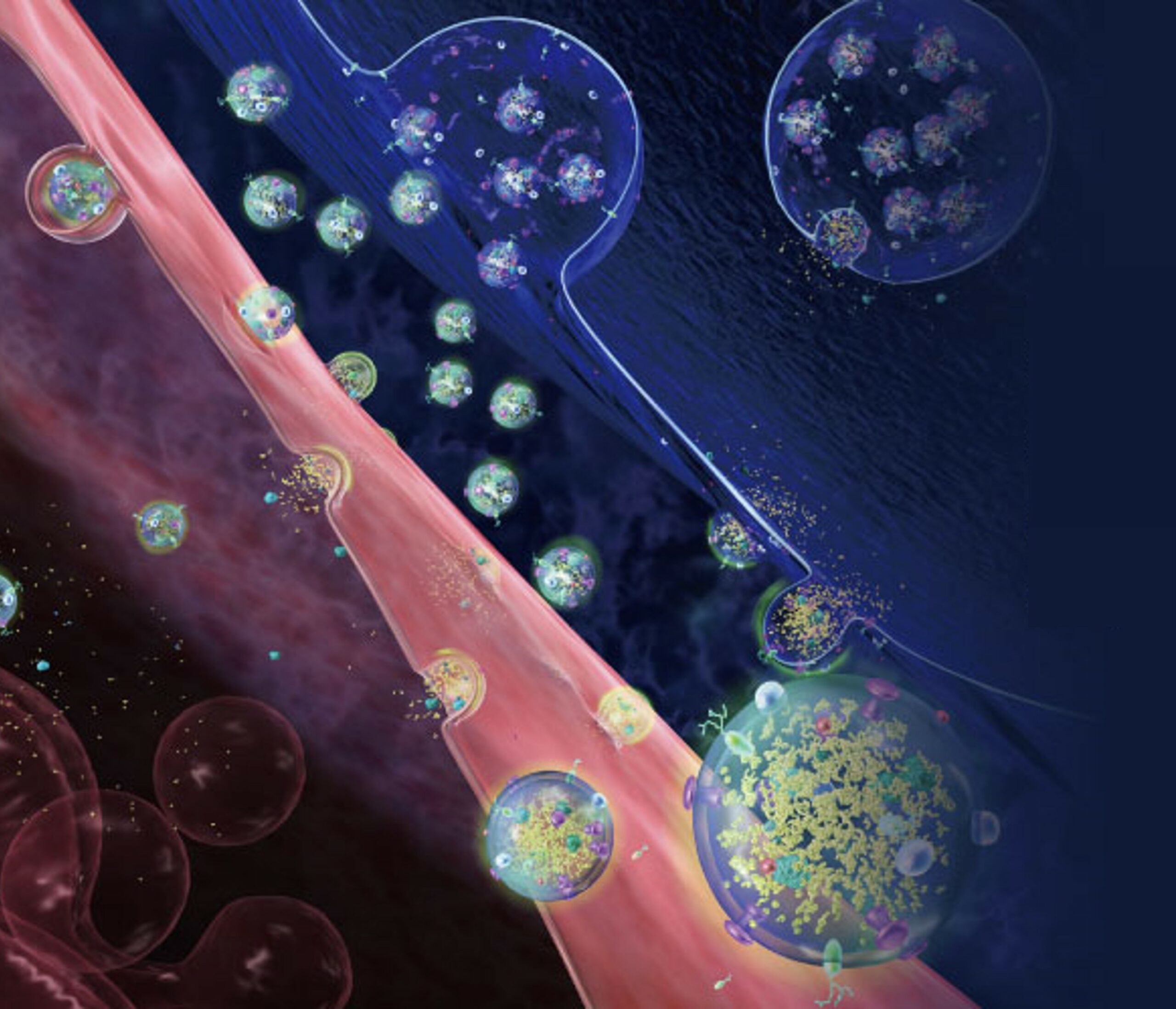
Over the course of the past ten years, EVs have attracted a growing amount of attention among the various liquid biopsy biomarkers. EVs are small membrane-bound particles that are secreted by all types of cells and carry various molecular cargoes, such as proteins, lipids, and nucleic acids. EVs can mirror the qualities of their cells of the beginning and can adjust the way of behaving of beneficiary cells through intercellular communication [2].
The first report of EVs in liquid biopsy was published in 2011 by Skog et al., who detected tumor-derived EVs containing mutant EGFRvIII mRNA in the plasma of glioblastoma patients [3]. Since then, numerous studies have explored the potential of EVs as liquid biopsy biomarkers for various types of cancer, such as prostate cancer [4], breast cancer [5], lung cancer, colorectal cancer, and pancreatic cancer. For instance, you can see extracellular vesicles as liquid biopsies for prostate cancer in the image below.
EVs outperform other liquid biopsy biomarkers in a number of ways, including:
- They are more steady and bountiful than cfNAs, which are effectively corrupted by nucleases in the course.
- They are able to carry a variety of biomolecules, including DNA, RNA, miRNA, lncRNA, methylation markers, proteins, and metabolites, which can provide a complete picture of the biology of a tumor.
- They can be isolated from blood, urine, saliva, cerebrospinal fluid, ascites, and pleural effusion, among other biological fluids.
- They are better at capturing the dynamics and heterogeneity of tumors than CTCs, which are uncommon and challenging to isolate and characterize.
Be that as it may, EVs likewise face a few difficulties for their clinical application as fluid biopsy biomarkers, for example,
- The absence of normalized strategies for EV disconnection, portrayal, and measurement.
- The heterogeneity and intricacy of EV populaces are gotten from various sources and subtypes.
- The interference with the analysis of tumor-derived EVs is caused by extracellular components and non-tumor-derived EVs.
- The requirement for EV biomarker validation in clinically relevant, large-scale prospective studies.
Regardless of these difficulties, EVs have shown extraordinary commitment as liquid biopsy biomarkers for early malignant growth determination, guess expectation, treatment choice, and checking. EV-based liquid biopsy is anticipated to become a powerful tool for precision oncology in the near future as new technologies and platforms for EV detection and analysis, such as nanotechnology-based methods, single-EV analysis, and multiplexed assays, are developed.
Celer Diagnostics solution
Providing kits for optimal and quantitative EVs separation.
Providing specific and registered markers to identify breast cancer and clone cancer.
- Extracellular Vesicle-DNA: The Next Liquid Biopsy Biomarker for Early Cancer Diagnosis? – PubMed (nih.gov)
- Extracellular vesicles as a source of prostate cancer biomarkers in liquid biopsies: a decade of research | British Journal of Cancer (nature.com)
- Extracellular Vesicle–Based Multianalyte Liquid Biopsy as a Diagnostic for Cancer | Annual Review of Biomedical Data Science (annualreviews.org)
- Extracellular Vesicle-Based Multianalyte Liquid Biopsy as a Diagnostic for Cancer – PubMed (nih.gov)
- Circulating tumor cells and extracellular vesicles as liquid biopsy markers in neuro-oncology: prospects and limitations | Neuro-Oncology Advances | Oxford Academic (oup.com)